

Posted by Pure Dental | Nov 11, 2026
Hearing that a severely damaged or infected tooth might need extraction can feel overwhelming. Losing a natural tooth can affect chewing, bite alignment, and confidence. Fortunately, advances in root canal treatment have created more opportunities to preserve teeth that once seemed impossible to repair. Microscopic endodontics uses powerful magnification and focused lighting to reveal tiny structures inside a tooth. This technology helps clinicians identify hidden canals, examine cracks, and treat complex infections with greater precision.
At Pure Dental, Dr. Vaughn emphasizes careful evaluations and patient-centered dental care. Before deciding whether extraction is necessary, understanding available tooth-saving treatments can help patients make informed decisions. For residents dealing with persistent tooth pain, consulting our dentist in Lake Ridge provides an opportunity to investigate the problem and explore appropriate treatment options.
What Is Microscopic Endodontics in Dentistry?
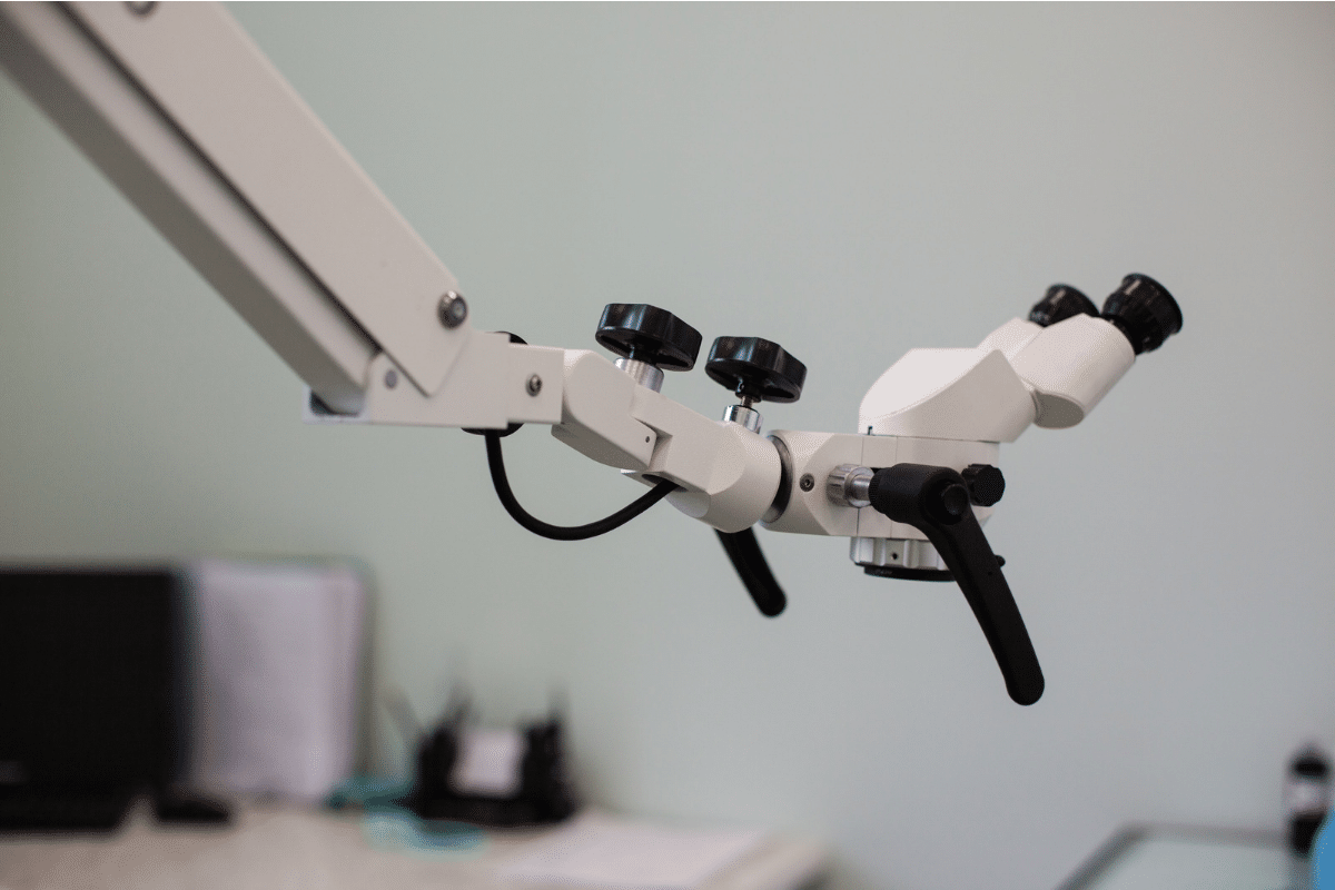
Microscopic endodontics is an approach to root canal treatment that uses a dental operating microscope to magnify a tooth’s internal structures.
According to the American Association of Endodontists (AAE), operating microscopes can provide magnification up to 25 times greater than unaided vision.
Traditional root canal treatment relies on clinical examination, imaging, instruments, and direct visualization. Microscopes add powerful illumination and magnification, allowing clinicians to examine areas that may otherwise remain difficult to see.
This increased visibility is particularly valuable when treating narrow canals, complex root anatomy, or previously treated teeth.
Although microscopes improve precision, treatment outcomes also depend on infection control, tooth condition, and proper restoration.
Why Severely Damaged Teeth Face Extraction
A tooth may require removal when damage prevents it from functioning safely or supporting a lasting restoration.
However, some teeth initially considered difficult to preserve may remain treatable after a thorough examination.
Common concerns include:
- Deep tooth decay: Bacteria reach the pulp and cause inflammation or infection.
- Untreated root canal infection: Infection spreads into tissues surrounding the root.
- Cracked tooth structure: Fractures threaten the tooth’s strength and stability.
- Failed previous treatment: Bacteria remain inside untreated canals or reenter through damaged restorations.
- Complex root anatomy: Narrow, curved, or calcified canals make treatment more challenging.
High-powered optical technology helps clinicians investigate these problems before determining whether extraction is necessary.
How Dental Microscopes Help Preserve Teeth
A dental operating microscope does more than enlarge the treatment area. It helps clinicians recognize structural details that influence treatment decisions.
Finding Hidden and Narrow Root Canals
Teeth contain intricate canal systems that vary in shape and number. Some molars have additional canals that can be difficult to identify.
If infected tissue remains inside an untreated canal, infection may persist after root canal therapy.
Magnification helps reveal these hidden pathways, allowing more thorough cleaning and disinfection.
Detecting Small Cracks and Fractures
Some tooth cracks remain difficult to see during routine examinations. Powerful lighting and magnification can help identify their location and extent.
This information supports decisions about whether a tooth can receive root canal treatment and a protective restoration.
However, a vertical root fracture or an extensive split may still make extraction necessary.
Preserving Healthy Tooth Structure
Removing unnecessary tooth material can weaken an already compromised tooth.
Microscopic visualization supports precise access to root canals while limiting avoidable removal of surrounding dental tissue.
Preserving sound tooth structure can improve the tooth’s ability to support its final restoration.
Managing Complicated Root Canal Problems
High-powered optics can also help remove broken instruments, identify canal blockages, and repair certain perforations. These procedures require careful case selection. Not every complication can be resolved through magnification alone.
Can Microscopic Treatment Save Previously Treated Teeth?
A tooth that develops pain or infection after root canal therapy does not automatically require extraction. Endodontic retreatment may offer another opportunity to preserve the tooth.
During retreatment, a clinician removes previous filling material, examines the canal system, and addresses any remaining infection.
Microscopes can help locate canals missed during the original procedure or identify other problems that may contribute to treatment failure.
If you have persistent discomfort after previous treatment, visit our dental office for an evaluation.
Our trusted dentist in Lake Ridge can examine the affected tooth and discuss whether retreatment, additional imaging, or referral for advanced endodontic care is appropriate.
What Happens During Microscope-Assisted Root Canal Treatment?
The exact procedure depends on the tooth’s condition and whether initial treatment or retreatment is necessary.
A typical process includes:
- Detailed examination: Clinical testing and dental imaging help identify infection, fractures, and remaining tooth structure.
- Local anesthesia: The treatment area is numbed to support patient comfort.
- Tooth isolation: A dental dam helps protect the treatment area from saliva and bacteria.
- Magnified canal access: The clinician uses illumination and magnification to identify the canal openings.
- Cleaning and disinfection: Small instruments and irrigation solutions remove infected tissue and reduce bacteria.
- Sealing and restoration: The canals are filled and sealed, followed by a suitable filling or crown.
Microscope use varies by clinical need and provider. A detailed consultation can clarify which technology and treatment approach are available.
When Is Tooth Extraction Still Necessary?
Although microscopic endodontics can expand treatment possibilities, not every tooth can be preserved.
Extraction may remain appropriate when:
- A vertical root fracture severely compromises the tooth.
- Extensive decay leaves insufficient structure for restoration.
- Advanced gum disease causes significant loss of bone support.
- The tooth has a severe split or an unfavorable fracture pattern.
- Previous treatment complications cannot be predictably repaired.
The decision should consider long-term tooth survival, not simply whether the infection can be temporarily controlled.
Why Preserving Natural Teeth Matters
Your natural teeth help maintain chewing efficiency, speech, and comfortable bite alignment. Removing a tooth may create a gap that allows neighboring teeth to shift over time.
Replacement options, including dental bridges and implants, can restore function. However, these treatments involve additional procedures and considerations.
Whenever a tooth remains structurally restorable, preservation deserves careful consideration.
For patients in Lake Ridge, Woodbridge, and nearby Prince William County communities, early evaluation can prevent a manageable dental problem from becoming more complicated.
Explore Your Options Before Tooth Extraction
A recommendation for tooth removal does not always mean there are no alternatives. Microscopic endodontics may provide additional opportunities to address hidden canals, persistent infections, and certain structural problems.
At Pure Dental, Dr. Vaughn helps patients understand their oral health concerns and available treatment options.
If you need a trustworthy dentist in Lake Ridge, book an appointment at our Woodbridge office. We welcome patients from Lake Ridge, Woodbridge, and surrounding communities who want to explore ways to preserve their natural teeth.
Frequently Asked Questions
1. Can microscopic endodontics save a badly infected tooth?
Microscope-assisted treatment may help preserve infected teeth by revealing canals and anatomical details that are difficult to see. However, success depends on the infection, remaining tooth structure, and bone support. A clinical examination can determine whether root canal therapy is appropriate.
2. Is microscope-assisted root canal treatment painful?
Root canal procedures typically involve local anesthesia to numb the affected area. You may experience pressure during treatment and temporary tenderness afterward. Discussing discomfort and anxiety during a consultation helps our dental team plan measures to keep you comfortable.
3. Can a failed root canal be treated again?
Yes. Endodontic retreatment can sometimes resolve persistent infection after a previous root canal. Magnification may help identify missed canals or complications. An examination and appropriate imaging can determine whether retreatment offers a reasonable chance of preserving the tooth.
4. Does microscopic endodontics guarantee tooth preservation?
No. Magnification improves visibility but cannot reverse every type of structural damage. Severe fractures, extensive decay, or inadequate bone support may still require extraction. A professional evaluation is necessary to determine whether preserving the tooth is clinically realistic.
5. How long can a tooth last after root canal treatment?
A successfully treated and restored tooth can function for many years, sometimes for a lifetime. Longevity depends on restoration quality, oral hygiene, bite forces, and future decay. Regular examinations help identify concerns before they threaten the tooth.
6. Should I seek another evaluation before extraction?
If extraction has been recommended and the situation is not an emergency, seeking clarification about tooth-saving options can be valuable. An additional evaluation may identify whether retreatment or endodontic surgery is possible. Prompt assessment is especially important when pain or swelling develops.
**Disclaimer: This content should not be considered medical advice and does not imply a doctor-patient relationship.
More Blog Posts
Can Microscopic Endodontics Help Prevent Tooth Extraction?
Hearing that a severely damaged or infected tooth might need extraction can feel overwhelming. Losing a natural tooth can affect chewing, bite alignment, and confidence. Fortunately, advances in root canal treatment have created more opportunities to preserve teeth that once seemed impossible to repair. Microscopic endodontics uses powerful magnification and focused lighting to reveal tiny structures inside a tooth. This
Cavity Prevention Guide: What Every Parent in Dale City Should Know
Learn how to protect your child’s teeth in Dale City, VA. Dr. Vaughn shares brushing tips, diet advice, fluoride info, and sealant benefits. Book your visit today!
What Happens During a Filling: Each Step Explained Calmly
Wondering what happens during a filling? Dr. Vaughn at Pure Dental explains each step calmly for Lake Ridge patients. Book your visit today.
How Your Breathing Habits Affect Your Face and Energy Levels
Have you ever woken up feeling exhausted despite sleeping for eight hours? Or noticed that your child frequently sleeps with their mouth open? These everyday observations may reveal something important about breathing habits. The way you breathe can influence oral health, sleep quality, and facial development, particularly during childhood. At Pure Dental, Dr. Vaughn helps patients understand concerns affecting their
Single-Tooth or Full-Arch Implants – Which Is Best for Woodbridge Residents?
When it comes to dental implants, deciding whether to go for a single-tooth or full-arch implant is a crucial decision for many. Whether you are in the bustling suburban community or nearby, where daily routines and lifestyle choices impact your oral health, it’s essential to understand which option might be best for you. The climate, family gatherings, and the local